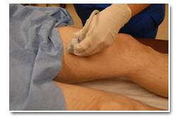
Vascular Ultrasound
Our portable ultrasound program employs only fully licensed, credentialed and qualified sonographers in accordance with Medicare IDTF guidelines. With our credentialed Ultrasound Sonographers, PPX offers the most extensive examination portfolio: including Doppler and testicular studies. Services available in select areas only. Contact PPX to learn if this service is available in your area.
Doppler ultrasound – used to see structures inside the body, while evaluating blood flow at the same time. Doppler ultrasound can determine if there are any problems within the veins and arteries.
Vascular ultrasound & ABI – used to see the vascular system and its function, including detection of blood clots. Our sonographers maintain required credentials for vascular studies, Registered Vascular Technologist, (RVT)
Lower Extremity Venous Duplex
Venous Doppler—used to see the venous system of the upper and lower extremities (to detect blood clots)
1. INDICATIONS
a. Phlebitis
b. Deep vein thrombosis (DVT)
c. Pulmonary embolus (PE)
d. Swelling
e. Extremity pain
f. Erythema
g. Cellulitis
2. PREPARATIONS
a. None
3. EXAM DESCRIPTION
The patient is examined in a supine position. The deep veins of the legs are examined with Spectral Doppler and Color Flow Imaging to evaluate for vascular patency and the presence or absence of thrombus and/or collateral vessels. Procedure time is 30 minutes per leg.
Ultrasound of the Carotids
Carotid—used to check the arteries of the neck for stenosis and/or blockage.
1. INDICATIONS
a. Carotid bruit
b. Dizziness
c. Syncope
d. Prior to major surgery, e.g., CAB, AAA
e. Follow up endarterectomy
f. Cerebrovascular accident (CVA)
g. Transient ischemic attack
h. Recurrent ASD
i. Aneurysm
j. Dissection
k. Carotid body tumor
l. Amaurosis fujax
m. Trauma
2. PREPARATIONS
a. None
3. EXAM DESCRIPTION
Patient is examined in the supine position. Imaging and Doppler Spectral Analysis are performed on the carotid, subclavian and vertebral arteries bilaterally. From the images the radiologist will give an estimate of the present stenosis as well as a characterization of any atheromatous plaque seen. Color Doppler Imaging is also utilized to further evaluate the presence or absence of hemodynamic flow abnormalities. Procedure time is approximately 30-45 minutes.
Arterial Doppler
Arterial Doppler—used to see the arterial system of the upper and lower extremities (to detect stenosis and/or blockage)
Patient Preparation- Patient will be asked to lie in bed. The extremity that is to be evaluated must be bare. (i.e. if the right lower extremity is to be evaluated the patient’s pants must be removed).
ABI—Ankle Brachial Index—done to screen for peripheral arterial disease-this test is done in conjunction with an arterial Doppler of the affected extremity. This exam is done by taking blood pressures of both of the ankles and both of the arms
Patient Preparation-Patient will be asked to lie in bed. Blood pressure cuffs will be placed on each upper arm to obtain arm blood pressures and will be placed on each calf to obtain ankle pressures.